Advanced Histopathology Services for Accurate Diagnosis.
Comprehensive Analysis for Early Detection, Diagnosis, and Treatment Planning.
What is Histopathology?
Histopathology is the study of tissue samples under a microscope to diagnose various diseases, especially cancer, infections, and inflammatory conditions. It plays a pivotal role in identifying abnormal cell structures, allowing clinicians to accurately plan treatments.
Why is Histopathology Analysis Important?
Histopathological analysis is essential for:
- Accurate Diagnosis
- Early Detection
- Customized Treatment Plans
- Prognosis Evaluation
- Informed Clinical Decisions
- Continuous Monitoring
Our Histopathology Process
Tissue Collection
Proper sample collection is crucial for accurate diagnosis. We provide clear instructions to ensure quality samples are obtained
Laboratory Examination
The tissue sample is processed and examined using advanced microscopes and staining techniques to identify abnormalities
Report Generation
Our expert pathologists generate detailed, easy-to-understand reports, ensuring timely delivery for optimal clinical decision-making
Work Flow
Here are key process steps we follow in our services:
1. Specimen Generation : Once doctors perform diagnostic or therapeutic surgical procedures on patients, tissue specimens are generated at healthcare facilities such as hospitals or clinics. Occasionally cytology and hematology specimens that are generated at healthcare facility are processed and prepared slides are sent to us for reporting.
2. Test request form generation : A detailed test request form filled with patient data, clinical and procedure description duly signed by concerned physicians are kept ready for transportation.
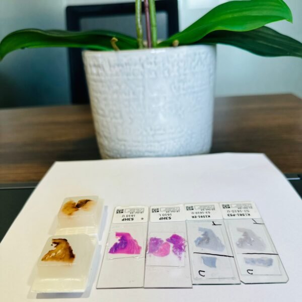
3. Specimen Fixation : Once biopsy or surgical specimens are obtained from a patient, the samples are properly labelled and kept for fixation.
4. Sample Transportation : Tissue specimens in proper fixative media or tissue slides in case of second opinion/ cytology/ hematology are properly packed, labelled and sent to us for further processing.
5. Grossing : Once the surgical tissue is received it is adequately sampled during grossing & representative tissue is processed for microscopy.
6. Tissue Processing : The tissue undergoes processing, including dehydration, clearing, and embedding in paraffin wax.
7. Tissue Microtomy : Thin sections of the embedded tissue are cut and placed on glass slides.
8. Staining : The tissue sections are stained using various techniques to highlight specific structures and cellular components.
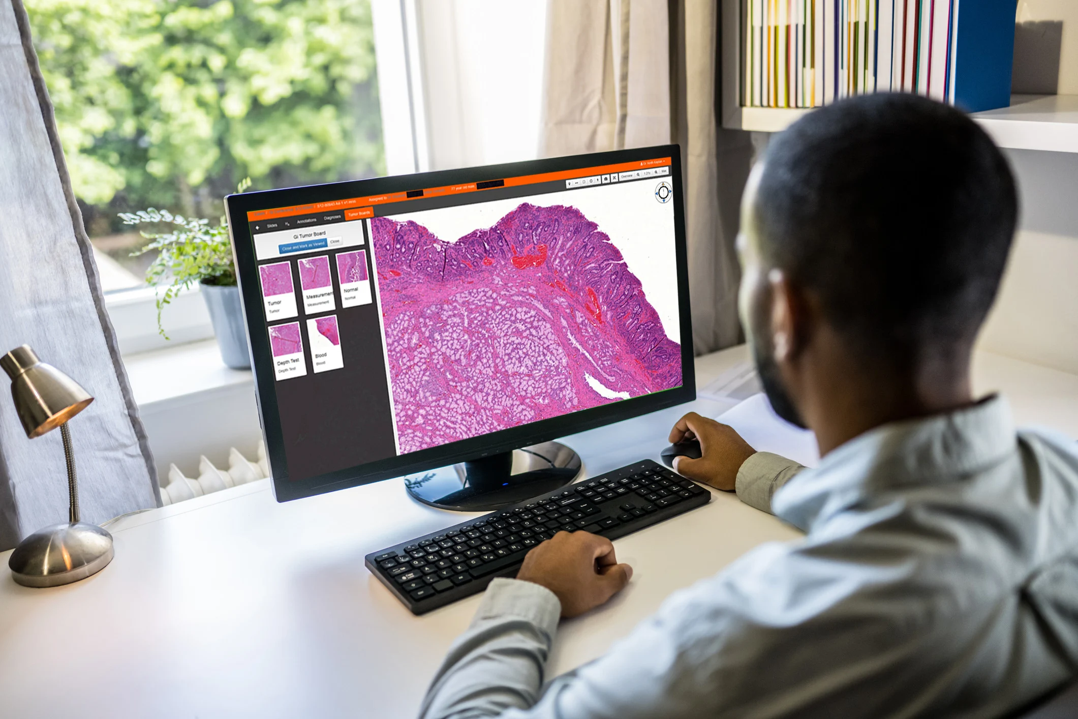
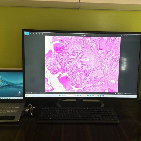
9. Digital Imaging : The prepared slides are scanned to create high-resolution digital images, which may include both macroscopic and microscopic views.
10. Image Transfer : These digital images, along with relevant patient information are securely transmitted to a remote telepathology facility.
11. Data Encryption : To protect patient privacy, data is encrypted during transmission and storage.
12. Image Reception : The telepathologist at the remote facility receives the images and verify their quality and completeness.
13. Image Analysis : The telepathologist examines the digital images, analyzes the tissue structure and cellular feature to make a diagnosis.
14. Diagnostic Report : A comprehensive diagnostic report is generated, including findings, impressions and in some cases recommendations for further studies and treatment.
15. Quality Assurance : The report undergoes a quality control process, which may include peer review, to ensure accuracy and compliance with standards.
16. Report Delivery : The final diagnostic report is securely transmitted back to the referring healthcare provider, enabling them to make informed decisions about patient management and treatment.
Why Choose Us?
• Experienced Professionals: Our team of skilled histopathologists and lab technicians are committed to providing accurate and timely results.
• State-of-the-Art Technology: We utilize the latest in diagnostic equipment and techniques to ensure precise tissue analysis.
• Quick Turnaround Time: We understand the importance of timely results, and ensure our clients receive reports without unnecessary delays
• Second Opinion Digital Pathology: At our second opinion digital pathology reporting service, we are your trusted resource for confirming and supporting medical diagnosis. Our team of expert pathologists use cutting-edge technology to review and analyze histopathology images and tissue samples, providing valuable second opinion. We empower doctors to make appropriate decisions about their patient management plan. We are here to add clarity and confidence in your patient treatment plan.
• Quality Assurance on Digital Pathology: As part of quality assurance service for pathology reporting, we have prioritized adherence to standardized protocols. Our dedicated team of expert pathologists meticulously review and validate every report and image to ensure the highest standard of accuracy and reliability. We employ rigorous quality control processes including IQA and EQA to guarantee that each diagnosis is precise and trustworthy. We have quality validation system in every step of tissue processing and reporting to uphold the quality of the medical reports, thereby providing the assurance of excellence .
Frequently Asked Questions (FAQs)
Histopathology once the biopsy is obtained from the patient this should be placed in 10% farmalinein an air tight / leak proff container. Once we are informed this will be collected.
Our typical turnaround time for histopathology reports is 2 days (urgent specimens), Biopsys ( 48-72 hours) , Resections 3-5 working days, depending on the complexity of the case.
Histopathology can be used to diagnose cancers, infections, autoimmune diseases, and other pathological conditions affecting tissues.